Plaat III
Table III

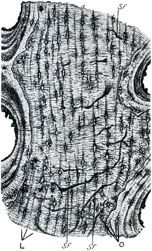
Afb. 5. Deel van de dwarsdoorsnede van een kuitbeen. (160 ×) L. streping door lamellen veroorzaakt, O. osteocyten, SF. vezels van Sharpey. De zeer fijne lijntjes zijn radiair verlopende canaliculi. - Uit: Maximow-Bloom, A textbook of histology. 7e dr. blz. 135. 1957. - Brief 90[49], blz. 22 en 26.
Ill. 5. Part of the cross-section of a fibula. (160 ×) L. striation caused by lamellae, O. osteocytes. SF. Sharpey's fibres. The very thin lines are canaliculi running in radial direction. - From: Maximow-Bloom, A textbook of histology. 7th ed. p. 135. 1957. - Letter 90[49], p. 23 and 27.

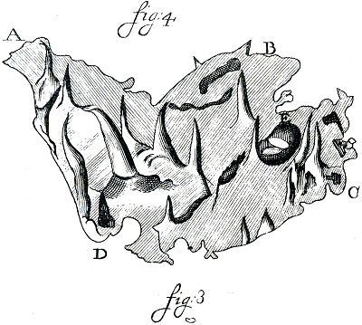
Fig. III. (L. 3). Stukje huid van de neus, natuurlijke grootte. - Brief 90[49], blz. 42.
Fig. IV. (L. 4). Afschilferend stukje huid van de neus. (± 60 ×) - l.c., blz. 42.
Fig. III. (L. 3). Part of the skin of the nose, natural size. - Letter 90[49], p. 43.
Fig. IV. (L. 4). Part of the skin of the nose, being peeled off. (± 60 ×) - l.c., p. 43.