Plaat XIII
Table XIII

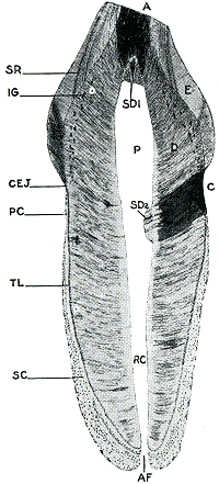
Afb. 14. Overlangse doorsnede van een snijtand van de mens. D. dentine met kanaaltjes, E. glazuur, P. pulpaholte, SC. cement met groeilijnen, SD1 en SD2. secundaire dentine. De zwarte plekken bij A en C zijn groepen luchthoudende dentinekanaaltjes als gevolg van resp. abrasie en cariës. - Uit: R. Kronfeld, Dental histology and comparative dental anatomy. blz. 37. pl. 4. 1943. - Brief 98[53], blz. 194, 204, 208.
Ill. 14. Longitudinal section of a human incisor. D. dentine with canaliculi, E. enamel, P. pulpal cavity, SC. cementum with incremental lines, SD1 and SD2. secundary dentine. The black spots at A and C are groups of dental canaliculi containing air, dead tracts, as the result of resp. abrasy and cariës. - From: R. Kronfeld. Dental histology and comparative dental anatomy. p. 37. table 4. 1943. - Letter 98[53], p. 195, 205, 209.

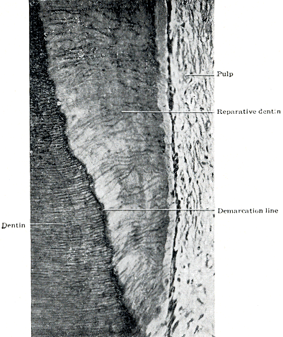
Afb. 15. Geslingerde kanaaltjes in secundaire dentine (reparative dentine). Zie ook kanaaltjes in normale dentine. - Uit: B.J. Orban, Oral histology and embryology. blz. 125. fig. 90. 1957. - Brief 98[53], blz. 194.
Ill. 15. Meandering canaliculi in reparative dentine. See also the canaliculi in normal dentine. - From: B.J. Orban, Oral histology and embryology. p. 125. fig. 90. 1957. - Letter 98 [53], p. 195

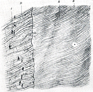
Afb. 16. Glazuur-dentinegrens. D. dentinekanaaltjes, aan het einde vertakt, S. glazuur met glazuurprisma's. - Uit: P. Stöhr, Lehrbuch der Histologie und der mikroskopischen Anatomie des Menschen. blz. 333. fig. 328. 1951. - Brief 98[53]. blz. 194-198.
Ill. 16. Border of enamel and dentine. D. dental canaliculi, branched at the end, S. enamel with enamel-prismata. - From: P. Stöhr, Lehrbuch der Histologie und der mikroskopischen Anatomie des Menschen. p. 333. fig. 328. 1951. - Letter 98[53], p. 195-199.